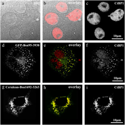
FIGURE 1.
Immunofluorescence of CtBP1 in COS-7 cells in the absence and presence of Bassoon. (a–c) DIC images (a) together with immunostainings against CtBP1 (c) of the same cells showed clear nuclear localization of CtBP1. (d–i) Disruption of the nuclear localization of CtBP1 (f and i) in the presence of a Bassoon construct similar to the full length, namely, Bsn95-3938 (d) as well as in the presence of Bsn1692-3263 (g). In the overlay images (e and h), GFP-Bsn95-3938 and Cerulean-Bsn1692-3263 are shown in green, whereas endogenous CtBP1 stained with Alexa 594 is shown in red. Yellow denotes the degree of colocalization of these proteins. In both cases, CtBP1 was enriched outside the nucleus in contrast to its original localization pattern.

