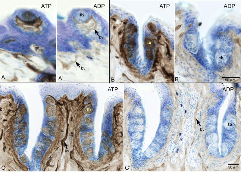
Figure 2.
Enzyme histochemical analysis revealing ecto-ATPase and ecto-ADPase activity via lead sulfide precipitate (brown) with thionin couterstain (blue) in taste buds in fungiform (A,A’), foliate (B, B’) and circumvallate (C, C’) papillae. With ATP as a substrate (A, B, C) strong ecto-ATPase activity is present in all taste buds (tb). Substituting ADP as a substrate results in lack of activity in the taste buds (A’, B’, C’) though some ecto-ADPase activity is present in nearby blood vessels (bv) (A’, C’). Scale bar in B’ also applies to A, A’, B. Scale bar in C applies to C’.

