Figure 5.

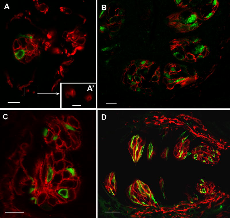
LSCM images of double label immunocytochemistry with NTPDase2 and the type II cell marker, PLCβ2 or the type III cell marker, 5HT. A, B: Staining for NTPDase2 (red) and PLCβ2 (green) in oblique sections cut through the fungiform (A) and foliate papillae (B). The NTPDase-immunoreactive cells are different from those exhibiting reactivity to either PLCβ2 or 5HT. NTPDase2 reactivity is also associated with nerve processes. Close observation of cross sections of this staining reveals that NTPDase2 immunoreactivity is limited to the periphery of the nerve profile while the core of the fiber remains void of staining, indicative either of membrane-associated neural reactivity or of glial cells circling the fiber (A’, scale bar 2.5μm) C, D: NTPDase2 (red) and 5HT (green) in foliate papillae (C) and cirumvallate papillae (D). Scale bars in A, B and C are 10μm; D scale bar is 25μm.
