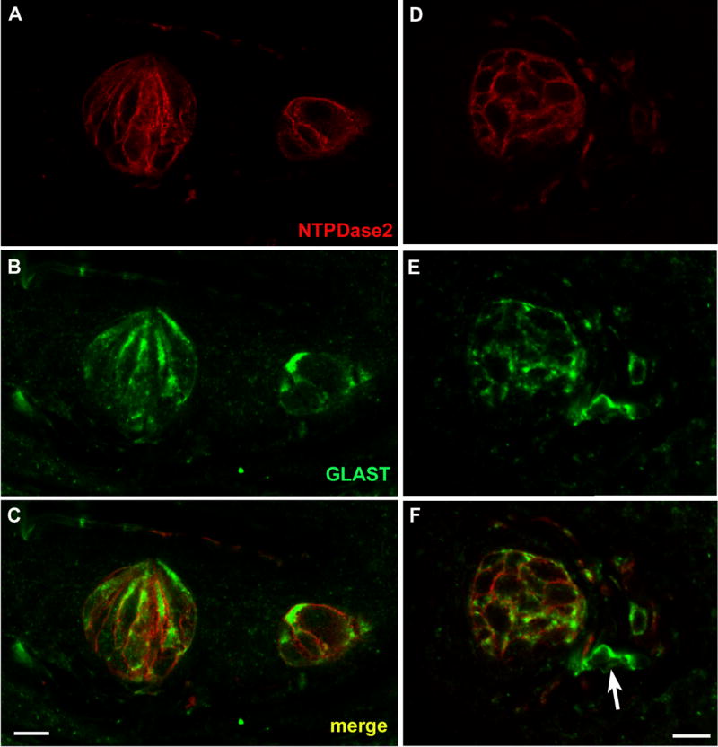
Figure 7.

LSCM images of double label assays with NTPDase2 and the type I cell marker, GLAST. A, D: NTPDase2 reactive cells in foliate and fungiform papillae, respectively. B, E: GLAST reactive cells in the same sections of foliate and fungiform papillae. C, F: Merged images of NTPDase2 and GLAST revealing colocalization of these markers to the same cellular membranes. The GLAST reactivity extends deeper into the cytoplasm than does the NTPDase2, and so appears as a green halo inside of the double-label (yellow) plasma membrane. GLAST reactivity is also associated with nerve fibers, though not all GLAST positive nerve fibers are double labeled with NTPDase2 as indicated with the arrow in F.
