
Figure 5.
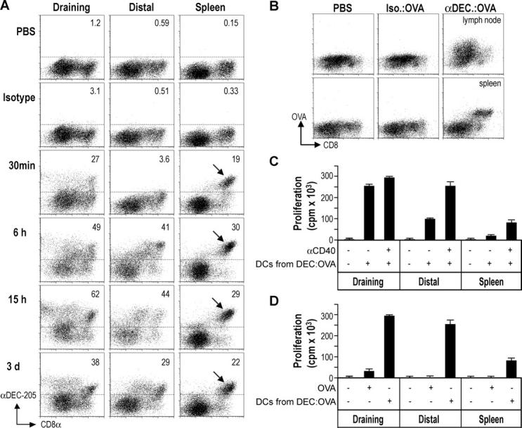
Systemic antigen presentation after DEC-205 targeting in situ. (A) C57BL/6 mice were given 10 μg of Alexa488-conjugated antibodies s.c. At the indicated time points, CD11c+ cells were enriched from the draining or distal lymph nodes or spleen for evaluation by flow cytometry. The frequencies of DCs capturing the injected Igs are shown, and the DEC-205 and CD8 high subset of splenic DCs arrowed. (B) C57BL/6 mice were given 10 μg of αDEC-205:OVA, isotype:OVA, or PBS s.c. and, after 18 h, CD11c+ cells were enriched from draining or distal lymph nodes or spleen. The presence of OVA was evaluated by intracellular staining with Alexa488-conjugated αOVA and flow cytometry. (C) 15 h after s.c. treatment with 5 μg of αDEC-205:OVA or the isotype conjugate ± αCD40, CD11c+ lymph node or spleen DCs were selected and used to stimulate OT-I T cells without further addition of OVA. (D) As in C, but mice were treated with αCD40 and either αDEC-205:OVA (5 μg), OVA (500 μg), or PBS. Data are representative of at least two experiments.