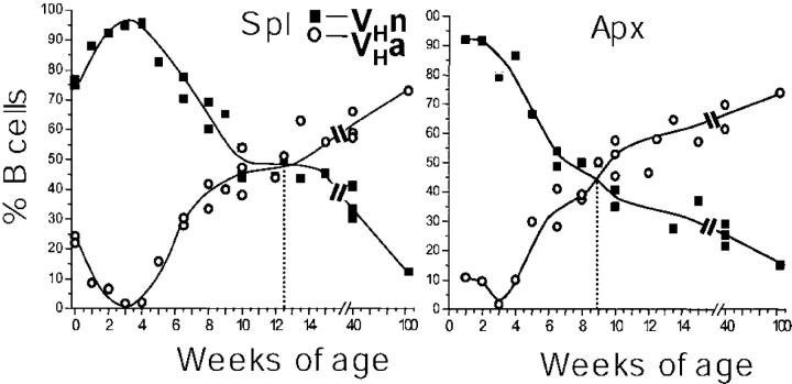
Figure 3.
Kinetics of the change in percentage of VHa and VHn B cells from birth to 2 yr of age in spleen and appendix of Alicia rabbits. Cells were stained with anti-IgM (FITC) and either anti-VHn (PE) or anti-VHa (PE) antibodies (see Materials and Methods), and the percentages of VHn (▪) and VHa (○) B cells for each rabbit are shown. The dotted line indicates the age at which VHa and VHn B cells each comprise 50% of the total number of B cells.

