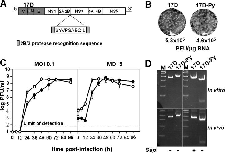
Figure 1.
Construction and viability of 17D-Py virus. (A) Schematic representation of P. yoelii CTL SYVPSAEQIL epitope insertion into the YF17D genome. The NS2B/NS3 protease recognition sequence was duplicated flanking the CTL epitope. (B) Infectious center assay from SW-13 cells transfected with 17D or 17D-Py RNAs. Plaques were fixed in 7.5% formaldehyde and stained by crystal violet. (C) One-step growth curve of 17D or 17D-Py in SW-13 cells infected at low (0.1 PFU/cell) or high (5 PFU/cell) multiplicity. Virus titers were measured by plaque assay on SW-13 cells as described in Materials and methods. The graphs represent the mean of three independent infections. (D) RT-PCR products of the 2B/3 region amplified from viral RNA recovered after infection of naive SW-13 cells with 17D or 17D-Py1 (in vitro) or from brain homogenate of suckling mice injected i.p. with each virus (in vivo). The fragments were digested with SspI and run on a 5% acrylamide gel. The P. yoelii CTL insert contains an SspI site not present in 17D.

