
Figure 2.
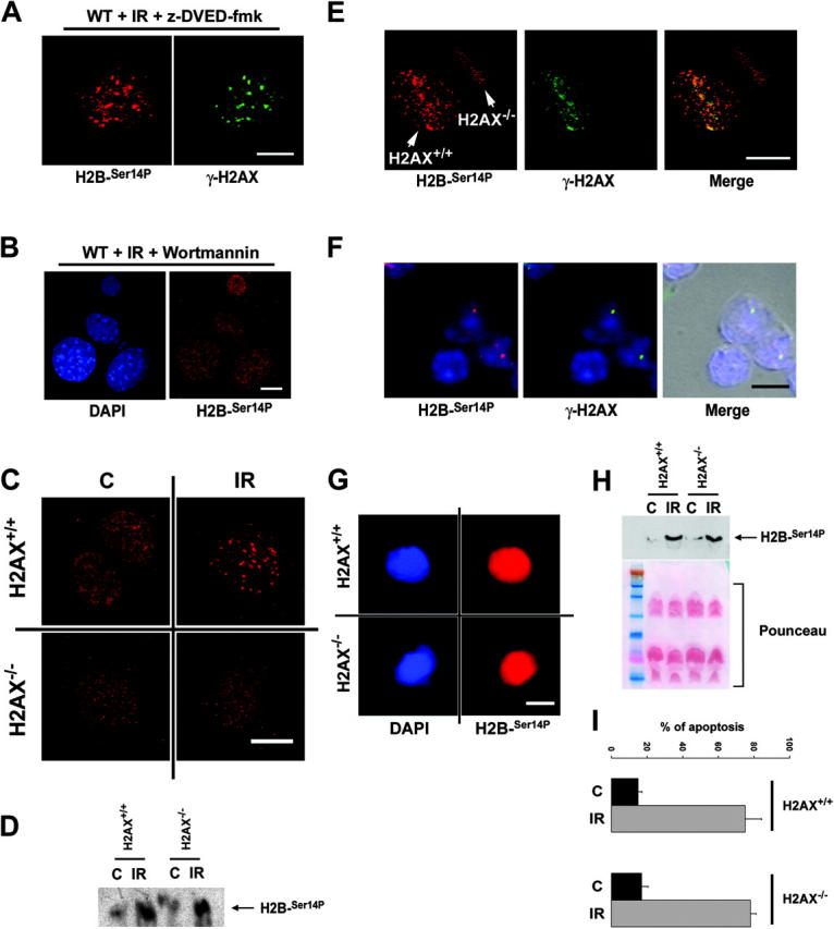
H2B-Ser14P IRIF formation is not associated with the apoptotic phosphorylation of H2B but is dependent on γ-H2AX. (A) Immunostaining of H2B-Ser14P in wild-type MEFs 4 h after treatment with 10 Gy of IR. The caspase inhibitor z-DEVD-fmk (200 μm) was added to the cells 1 h before the exposure to IR and was maintained in the medium throughout the length of the experiment. Bar, 10 μm. (B) Immunostaining of H2B-Ser14P in wild-type MEF 4 h after treatment with 10 Gy of IR. The PIKK inhibitor wortmannin (200 μm) was added to the cells 15 min before irradiation and was maintained in the medium throughout the length of the experiment. Bar, 10 μm. (C) Immunostaining of H2B-Ser14P (red) in H2AX+/+ and H2AX−/− MEFs 4 h after mock treatment (C) or exposure to 10 Gy of IR. Bar, 5 μm. (D) Western blot analysis of H2B-Ser14P levels in H2AX+/+ and H2AX−/− MEF 4 h after exposure to 100 Gy. (E) Immunostaining of H2B-Ser14P (red) and γ-H2AX (green) in a mixed population of H2AX+/+ and H2AX−/− MEFs that had been exposed to laser damage. Bar, 5 μm. (F) Immunostaining of H2B-Ser14P (red) and γ-H2AX (green) in freshly isolated thymocytes. Bar, 5 μm. (G) Example of the massive H2B-Ser14P (red) staining in H2AX+/+ and H2AX−/− apoptotic thymocytes (see H and I). Bar, 5 μm. (H) Western blot detection of H2B-Ser14P in histone extracts that were prepared by acid extraction from H2AX+/+ and H2AX−/− thymocytes 8 h after exposure to 5 Gy of IR. The Ponceau staining of the nitrocellulose membrane is shown as a loading control. (I) Apoptosis in irradiated thymocytes (IR) measured by flow cytometric analysis of the percentage of cells with a sub-G1 DNA content. Nonirradiated cells (C) that were kept in culture media during the 8-h period were used as a control. When shown (A, B, E, and F), DNA was counterstained with DAPI (blue).