Figure 2.
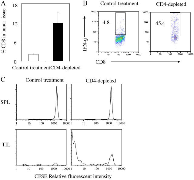
CD4+ cells suppress the proliferation and IFN-γ production of tumor-infiltrating CD8+ T cells. 106 Ag104Ld tumor cells were inoculated to C3B6F1 mice s.c. and anti-CD4 antibody (GK1.5) was injected i.p. 14 d after tumor challenge. Spleen as well as tumor tissue were isolated from mice 1 wk after CD4 depletion. The percentage of CD8+ T cells in the tumor tissue was determined by FACS (a). The tumor-infiltrating T cells (TIL) were enriched with anti-Thy1.2 magnetic bead system. Spleen cells and purified TIL were stimulated in vitro with PMA and ionomycin in the presence of brefeldin A for 4 h. The percentage of IFN-γ–producing CD8+ T cells among total CD8+ T cells in the tumor was determined by FACS (b). 106 MC57-SIY tumor cells were injected at multiple sites s.c. to 2C TCR transgenic mice in Rag-1 − / − background to activate 2C T cells. 2C T cells were isolated 96 h after activation and of CD62LloCD44high phenotype. CFSE-labeled activated 2C T cells were adoptively transferred to these Ag104Ld tumor bearing mice with or without CD4 depletion. Tumor tissue was collected from mice 2 d after transfer of 2C T cell. The proliferation of CFSE-labeled 2C T cells was monitored by FACS (c). Results from one experiment representative of three were shown.

