
Figure 5.
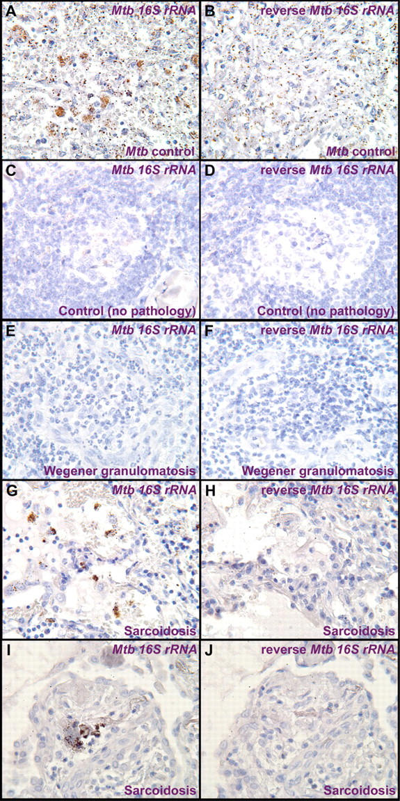
Cellular localization of Mtb 16S rRNA DNA by ISH. Shown are representative photomicrographs from biopsies of Mtb-infected tissue (A and B), normal controls (no pathology; C and D), Wegener granulomatosis (E and F), and sarcoidosis tissues (G–J). Tissue sections were hybridized with or without DIG-labeled probes, detected with anti-DIG F(ab′)2 fragments conjugated with alkaline phosphatase, and developed with Vector Red substrate. Mtb-infected tissue (A) and sarcoidosis samples (G and I) demonstrated focal collections of Mtb 16S rRNA DNA that were not seen in Wegener granulomatosis (E) nor in nongranulomatous control tissues (C). All tissues were negative using reverse Mtb 16S rRNA probes.