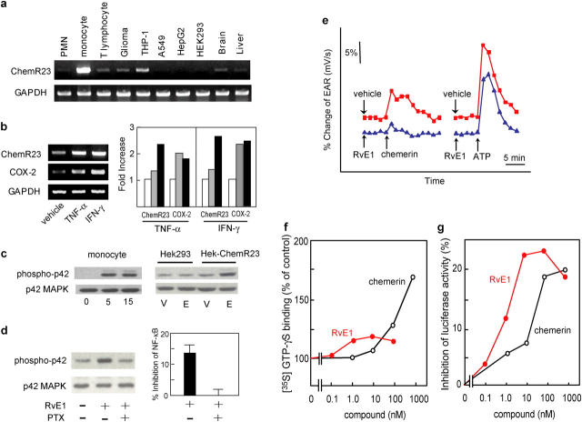
Figure 5.
Receptor expression. (a) RT-PCR analysis of human peripheral blood leukocytes and glioma (DBTRG-05MG), monocytic (THP-1), lung epithelial (A549), hepatoma (HepG2), embryonic kidney (HEK293) cell lines, and brain and liver. (b) RT-PCR analysis of human peripheral blood monocytes exposed to either buffer alone, 10 ng/ml TNF-α, or 25 ng/ml IFN-γ for 6 h (gray bars) and 24 h (black bars). Expression levels were quantified by a National Institutes of Health image, normalized by GAPDH levels, and expressed as fold increase over vehicle-treated cells. (c) MAP kinase activation in human peripheral blood monocytic cells were exposed to RvE1 for 5 or 15 min and HEK-ChemR23 cells were treated with 100 nM RvE1 (E) or vehicle (V). (d) Pertussis toxin (PTX) blocks RvE1-induced ERK activation and NF-κB inhibition in HEK-ChemR23 cells. (e) EAR with HEK-ChemR23 cells exposed to vehicle (red line), 100 nM RvE1 (blue line), 10 μM chemerin peptide, or 10 μM ATP using Cytosensor microphysiometer. These responses were observed from three separate experiments. (f) Actions of RvE1 (red) and chemerin peptide (black) on [35S]-GTP-γS binding to HEK293 cell membranes expressing human ChemR23. (g) Inhibition of TNF-α induced NF-κB luciferase activities by RvE1 (red) and chemerin peptide (black) on HEK293 cells expressing human ChemR23.

