
Figure 1.
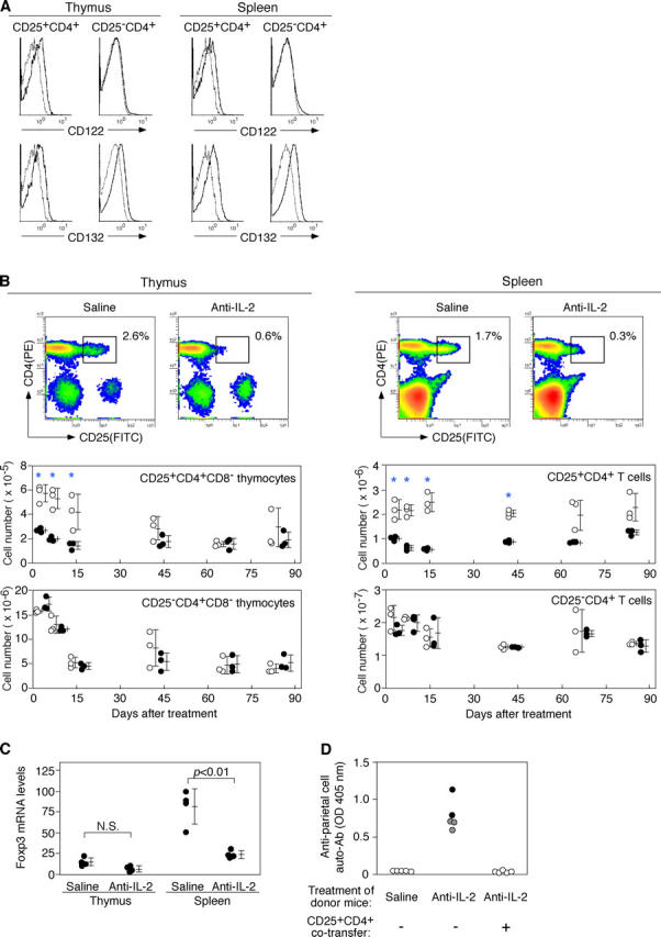
Neutralization of IL-2 by specific mAbs reduces the number of Foxp3 + CD25+ CD4+ T reg cells in normal naive mice. (A) Expression of CD122 (IL-2Rβ) and CD132 (IL-2Rγc) by CD25+ or CD25− CD4+ T cells in the thymus or spleen of normal BALB/c mice. Dotted lines represent control staining with isotype-matched mAb. (B) Representative flow cytometric profiles of thymus and spleen cells from 8-wk-old BALB/c mice 14 d after injection of anti–IL-2 mAb or saline. Kinetics of the number of CD25+ CD4+ T cells and CD25− CD4+ T cells after injection of anti–IL-2 mAb (black circles) or saline (white circles). Asterisks indicate significant differences (P < 0.05). (C) Relative Foxp3 mRNA levels assessed by real-time quantitative PCR with CD4+ CD8− thymocytes or CD4+ splenocytes from mice treated with anti–IL-2 mAbs. Quantity of Foxp3 normalized to HPRT. (D) BALB/c nude mice were i.v. transferred with spleen cells (3 × 10⁷) from BALB/c mice injected with anti–IL-2 mAbs or saline 14 d earlier with or without CD25+ CD4+ T cells (106) from nonmanipulated mice and examined histologically and serologically 3 mo later. Titers of anti-parietal cell autoantibodies were assessed by ELISA. Black circles, macroscopically and histologically evident grade 2 gastritis; gray circles, histologically evident grade 1 gastritis; white circles, intact gastritis mucosa. See reference 55 for histological grading.