Figure 1.
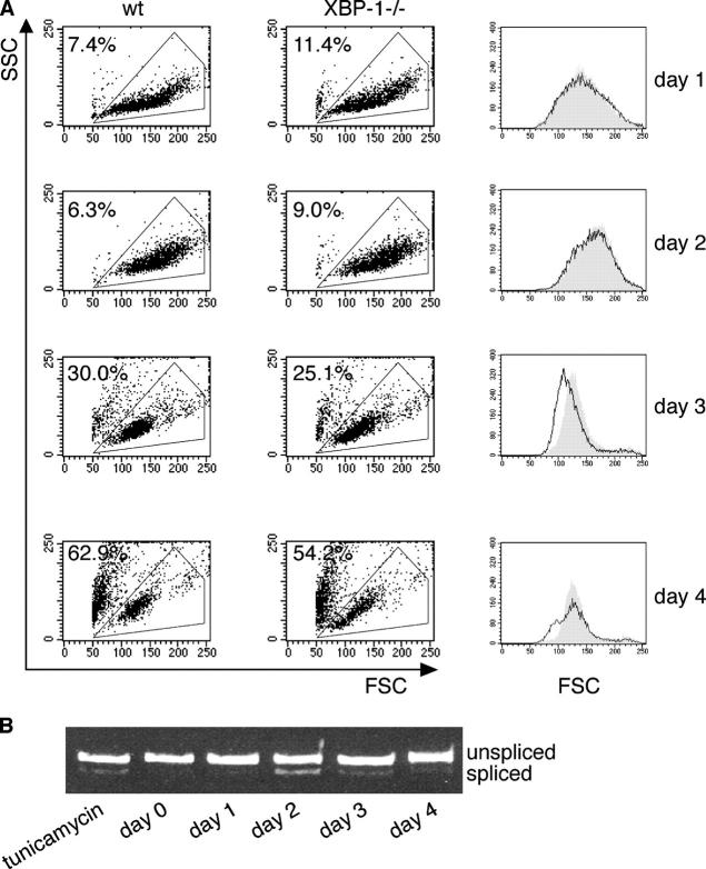
XBP-1 controls plasmablasts' cell size. (A) WT or XBP-1−/− B cells were purified from splenocytes by magnetic depletion with anti-CD43. Cells were plated at 106 cells/ml and stimulated with CpG. Flow cytometry analysis was performed every 24 h, and live cells were gated based on their forward and side scattering. Cells were replated at 106 cells/ml density and were analyzed the next day. Line graphs of the gated cells at the forward scatter channel (FSC) are shown in the right column. Gray, WT cells; white, XBP-1−/− cells. The percentage of dead cells is indicated. (B) Cells were stimulated with CpG for 4 d. RNA was extracted at the indicated times, and splicing of XBP-1 mRNA was analyzed by RT-PCR. Tunicamycin treatment (1μg/ml, 4 h) of naive B cells was used as a positive control.

