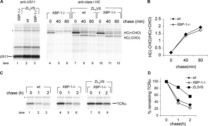
Figure 4.
Degradation of ER proteins in plasmablasts does not require XBP-1. (A) Cells stimulated 24 h days with CpG were transduced with a retrovirus that encodes HLA-A2-IRES-US11 and incubated in the presence of CpG. 2 d after infection, cells were pulse-labeled with [35S]methionine for 20 min and chased for up to 80 min in the presence or absence of the proteasome inhibitor ZL3VS. Cells were lysed in 1% SDS; the lysate was diluted to 0.07% SDS with NP-40 lysis mix followed by immunoprecipitation with anti-class I heavy chain serum (αHC) and analysis by SDS-PAGE (12%). US11 was immunoprecipitated sequentially from the zero time point and similarly analyzed. (B) Autoradiograms were quantified as mentioned above and the deglycosylated HC/glycosylated HC ratio was calculated. (C) Similarly to (A), cells were transduced with a retrovirus that encodes TCRα-IRES-YFP. Cells were pulse-labeled with [35S]methionine for 20 min and chased for up to 2 h in the presence or absence of the proteasome inhibitor ZL3VS. Cells were lysed in 1% SDS; the lysate was diluted to 0.07% SDS with NP-40 lysis mix followed by immunoprecipitation with anti-TCRα and analysis by SDS-PAGE (12%). (D) Autoradiograms were quantified as mentioned above and the relative amount of TCRα to the zero time point was calculated.

