
Figure 2.
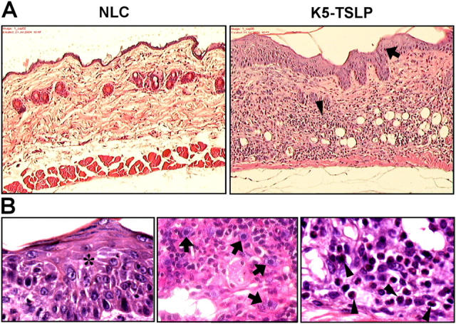
Histological observations of the skin in K5-TSLP mice. Paraffin-embedded sections of lesional skin from a K5-TSLP mouse and an NLC were stained with H&E. (A) Skin sections showing pronounced acanthosis (arrow) and dermal infiltration (arrowhead) at 10×. (B) Images of lesional skin from a K5-TSLP mouse showing epidermal spongiosis (asterisk) and dermal infiltration by mast cells (arrows) and eosinophils (arrowheads) at 100×.