Figure 5.
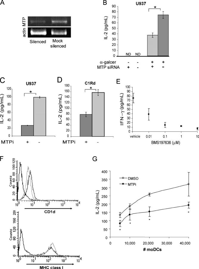
MTP in human cells is critical for CD1d antigen presentation. (A) U937 cells were treated with irrelevant or MTP-specific siRNA oligomers. RNA was isolated 48 h after silencing, and transcript levels of mtp and β-actin were determined by RT-PCR. (B) Silenced and mock-silenced U937 cells were incubated with α-galcer for 3 h, washed, and co-cultured with DN32 cells (E/T = 1:1). *, P < 0.001. Results are representative of two independent experiments. (C) U937 cells cultured with BMS212122 (MTPi) or vehicle for 3 d were incubated with α-galcer for 3 h, washed, and co-cultured with DN32 cells (E/T = 1:1). *, P < 0.001. (D) C1Rd cells cultured with BMS212122 (MTPi) or vehicle for 4 d were incubated with α-galcer for 4 h, fixed with 0.05% glutaraldehyde for 30 s, washed, and co-cultured with DN32 cells (E/T = 2:1). *, P < 0.02. Results are representative of three independent experiments. (E) C1Rd cells cultured with BMS197636 or vehicle were incubated with NKT cell lines derived from human peripheral blood in the presence of 1 ng/ml PMA. NKT cells co-cultured with C1R mock transfected cells in the same assay yielded 33.6 ± 9.8 pg/ml IFN-γ. (F) Human monocyte-derived DCs were stained with 42.1 (CD1d), anti–HLA-A,B,C (MHC class I), or isotype control antibodies (dashed lines) after 6 d of differentiation in the presence of BMS212122 (gray lines, MTPi) or vehicle control (black lines). Results are representative of two independent experiments. (G) Day 6 human moDCs differentiated in the presence of BMS212122 (○, MTPi) or vehicle (•) were incubated with α-galcer for 3 h, washed, and co-cultured with 50,000 DN32 cells. *, P < 0.05. Results are representative of two independent experiments. Values are ±SD.

