
Figure 1.
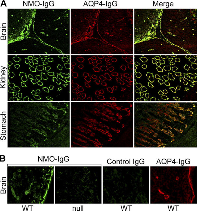
Immunofluorescence reveals NMO-IgG colocalization with AQP4 in mouse tissues. (A) Brain: Virchow-Robin space (pial–astrocyte interface) at the junction of two folia in mouse cerebellar cortex and midbrain. NMO-antigen (green, fluorescein-conjugated anti-human IgG), AQP4 water channel protein (red, rhodamine-conjugated anti–rabbit IgG); merged images yellow. Kidney: colocalization of NMO and AQP4 antigens in distal collecting tubules of medulla. Stomach: basolateral membranes of epithelial cells in deep gastric mucosa. (B) WT mouse brain binds NMO-IgG (panel 1) in a pattern that is indistinguishable from its binding of AQP4-specific IgG (panel 4); pia, subpia, and microvessels are stained. However, AQP4 knockout mouse brain (null) did not bind NMO-IgG (panel 2), and the serum of a control patient who had neuropsychiatric disease did not bind to WT brain (panel 3).