
Figure 6.
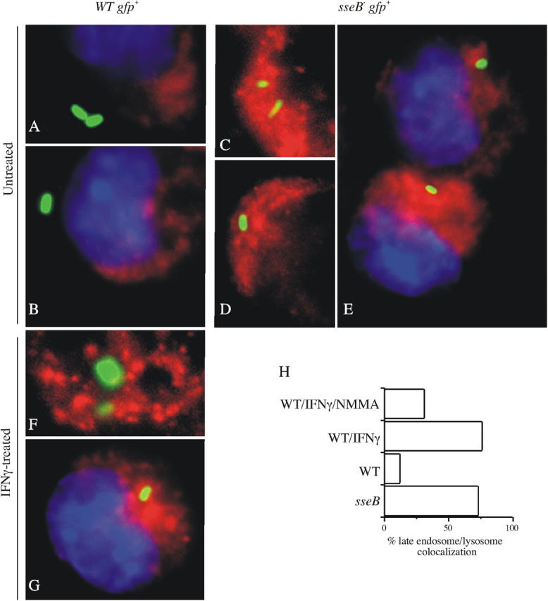
NO stimulates progression of the Salmonella phagosome along the degradative pathway. The intracellular localization of Texas red-dextran–labeled late endosomes/lysosomes (red), GFP-expressing Salmonella (green), and DAPI-labeled host cell nucleus (blue) was visualized by fluorescence microscopy. J774 cells were infected with either WT Salmonella (A and B) or its isogenic sseB mutant control (C–E). Selected groups of J774 cells were treated with IFNγ 20 h before infection with WT Salmonella (F and H). Samples were prepared for immunofluorescence microscopy 20 h after Salmonella infection and after 1 h of pulsing with Texas red-dextran. The percentage of Salmonella colocalizing with late endosomes/lysosomes is shown in H. These data represent an analysis of 330 independent observations from five separate experiments.