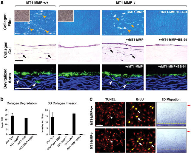
Figure 3.
Collagenolytic activity of isolated MT1-MMP−/− VSMCs. (a) Isolated WT cells, MT1-MMP-null VSMCs, or MT1-MMP–transduced null cells (rMT1-MMP) were cultured atop a film of type I collagen (upper row) or a 3-D gel of type I collagen (middle row) in the absence or presence of BB-94. Upper row: yellow arrowheads mark zones of collagen proteolysis. Insets are phase contrast micrographs of MT1-MMP+/+ and MT1-MMP−/− VSMCs that were cultured atop the collagen film substratum. Middle row: black arrows indicate the position of VSMCs that have invaded the collagen gels. Bottom row: fluorescently labeled VSMCs (green) were cultured atop devitalized aorta. White arrowheads mark the position of VSMCs that invaded the aortic tissue. Bars, 50 μm. (b) Quantitative analysis of the MT1-MMP–dependent collagenolytic and invasive activities displayed by smooth muscle cells cultured as described. Results are expressed as the mean ± SEM (n = 5). (c) VSMC proliferation (BrdU) and apoptosis (TUNEL) in WT and MT1-MMP−/− cultures established atop type I collagen gels. TUNEL-positive VSMCs (arrowheads) and BrdU-labeled VSMCs (arrows) are shown with propidium iodide counterstaining (red) used to visualize cells. Bar, 50 μm. Right panels: motility of MT1-MMP+/+ and MT1-MMP−/− VSMCs across a type I collagen–coated substratum. The dashed yellow line marks the position of cells at the start of the assay; the red arrow indicates the position of the leading front of cells after 72 h in culture. Bar, 0.5 mm.

