
Figure 4.
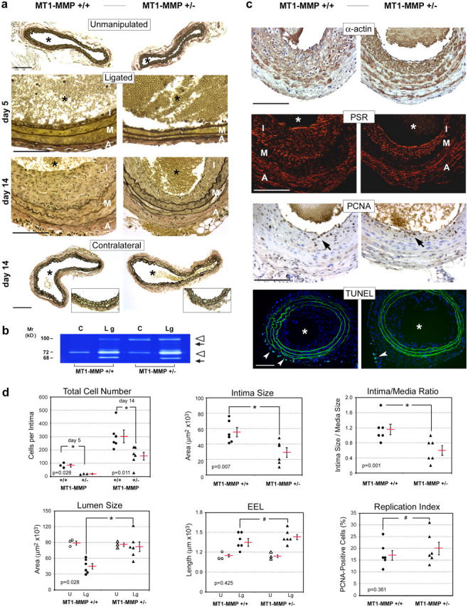
MT1-MMP deficiency reduces neointima formation in vivo. (a) Verhoeff Van Gieson's staining of unmanipulated controls as well as contralateral and ligated common carotid arteries from MT1-MMP+/+ and MT1-MMP+/− mice 5 d and 14 d after ligation. Elastic fibers are stained black; nuclei are stained brown. Bottom row insets demonstrate intact intima in contralateral carotids of either genotype. *, vessel lumen; A, adventitia; I, intima; M, media. (b) Gelatin zymography of carotid artery extracts 14 d after ligation. The pro- (open arrowheads) and processed (arrows) forms of MMP-9 (upper arrowhead and arrow) and MMP-2 (lower arrowhead and arrow) are indicated. C, contralateral artery; Lg, ligated artery. (c) 14 d after ligation, paraffin sections of ligated carotid arteries were probed with anti-smooth muscle α-actin polyclonal antibody (α-actin; brown stain) or stained with Picro-Sirius red (PSR; red stain). Cell proliferation and apoptosis were determined with anti-PCNA mAb (PCNA) and TUNEL, respectively. PCNA-positive nuclei (brown, arrows) and TUNEL-positive cells (arrowheads) are shown. (d) Quantitative assessment of vascular remodeling. Charts show individual numbers (six for each group) with the mean indicated by a red bar ± SEM. All data were obtained at day 14 after ligation except for total cell numbers, for which values are shown for both days 5 and 14. EEL, external elastic lumina. Bars, 100 μm.