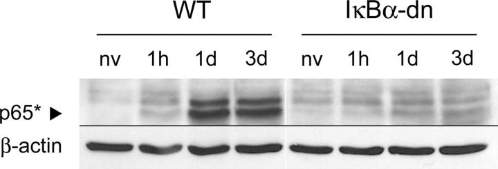
Figure 2.
Time course of NF-κB activation after SCI in WT and GFAP-IκBα-dn mice. NF-κB activation was evaluated by Western blot analysis with an antibody that specifically recognizes the activated form of p65 (p65*). The arrowhead indicates p65* migrating at exactly 65 kD. 20 μg of proteins/sample were loaded, and blots were probed for β-actin as a control. A representative blot is shown (n = 3). nv, naive.

