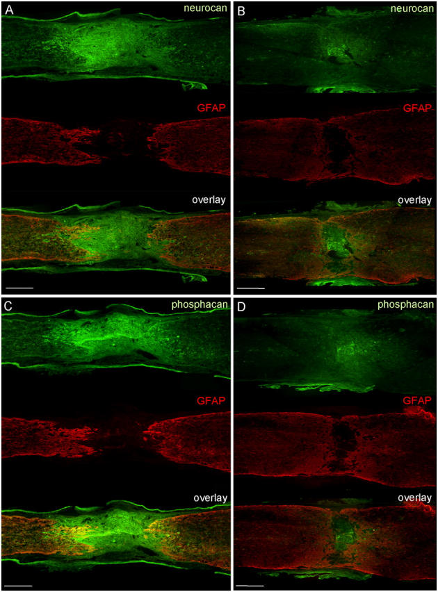
Figure 5.

Neurocan and phosphacan immunohistochemistry 8 wk after SCI. WT (A and C) and GFAP-IκBα-dn (B and D) sections of the spinal cord were double labeled for GFAP (red) and either neurocan (green; A and B) or phosphacan (green; C and D). Bars, 450 μm.
