
Figure 2.
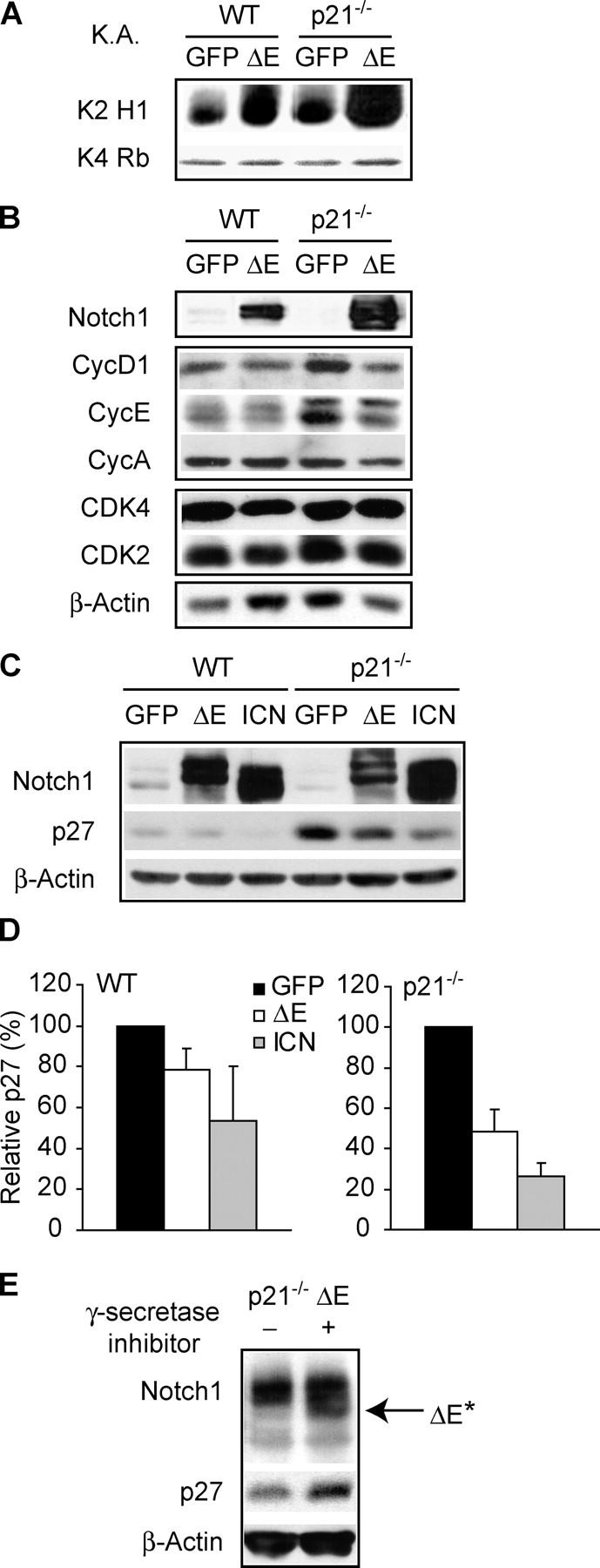
N1 activation correlates with increased CDK2 kinase activity and promotes p27Kip1 down-regulation. (A) In vitro kinase activity assay (K.A.) of immunoprecipitated CDK2 (K2) or CDK4 (K4) complexes from growing cells. Histone (H1) or retinoblastoma (Rb) was used as substrate, respectively. (B) Immunoblot of cyclins and CDKs. (C) Immunoblot of p27Kip1. p21−/− cells express higher levels of p27Kip1 compared with WT, perhaps as a compensatory mechanism. In this blot, where WT and p21−/− cells are side by side, p27Kip1 in WT samples is underexposed to avoid its overexposure in p21−/− samples. (D) ImageQuant densitometric analysis of p27Kip1 protein expression. The values, an average of five independent experiments, indicate percentage of p27Kip1 protein detected in ΔE or ICN cells relative to their GFP controls (100%). (E) Cells were incubated with the GSI (+) or DMSO vehicle control (−) for 12 h. Protein extracts from harvested cells were analyzed by Western blot to assess N1 and p27Kip1 expression.