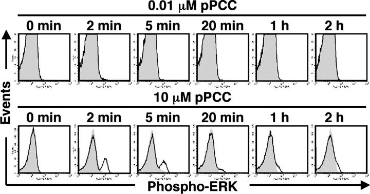
Figure 6.
Intense phosphorylation of ERK induced by high concentrations of peptide. Line 94 CD4+ T cells were pretreated with U 0126 (3 μM) or DMSO and stimulated with P13.9 cells that were preloaded with 0.01 or 10 μM pPCC. Cells were fixed at indicated time points, and intracellular staining for phospho-ERK was performed. The levels of phospho-ERK in U 0126- and DMSO-pretreated cells are shown in shaded and open line graphs, respectively. The experiment was carried out three times with consistent results.

