Figure 2.
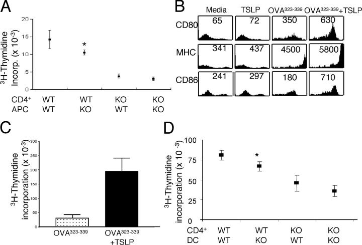
TSLP activates murine DCs. (A) In vitro antigen recall response. CD4+ T cells and APCs from immunized WT and TSLPR KO mice were cultured with 200 μg/ml of OVA for 48 h and then pulsed with [3H]thymidine. Shown are means ± SEM for seven experiments. (B) Splenic CD11c+ cells were sorted from the spleens of WT BALB/c animals and incubated overnight with media, TSLP alone, 5 μg/ml of OVA323–339 peptide, or with OVA323–339 peptide plus TSLP. TSLP treatment increased the surface levels of CD80, MHC class II, and CD86. Numbers represent mean fluorescent intensity. (C) Splenic DCs were incubated with 5 μg/ml of OVA323–339 peptide alone or with TSLP before being washed, treated with mitomycin C, and cultured with purified CD4+ T cells from DO11.10 RAG2−/− mice at a 1:10 ratio. Antigen presentation of DCs was enhanced significantly by TSLP treatment (P = 0.005, shown are means ± SEM for five experiments). (D) CD4+ T cells and DCs were purified from DO11.01/WT and DO11.10/TSLPR KO unmanipulated mice, and these were cultured together in the indicated combinations at a ratio of 1:10 DC/T cell, with 5 μg/ml of OVA323–339 peptide, and [3H]thymidine incorporation determined at 48 h. Replacing WT APCs with TSLPR KO-derived DCs moderately, but significantly, reduced the antigen-driven proliferation of DO11.10 Tg/WT CD4+ T cells (P = 0.04). WT DCs provided no significant enhancement over the weak expansion of DO11.10/TSLPR KO CD4+ T cells. Shown are means ± SEM for seven experiments. (A and D) Asterisks indicate that the difference between WT CD4+/WT APCs or DCs and WT CD4+/KO APCs or DCs was statistically significant (P < 0.05).

