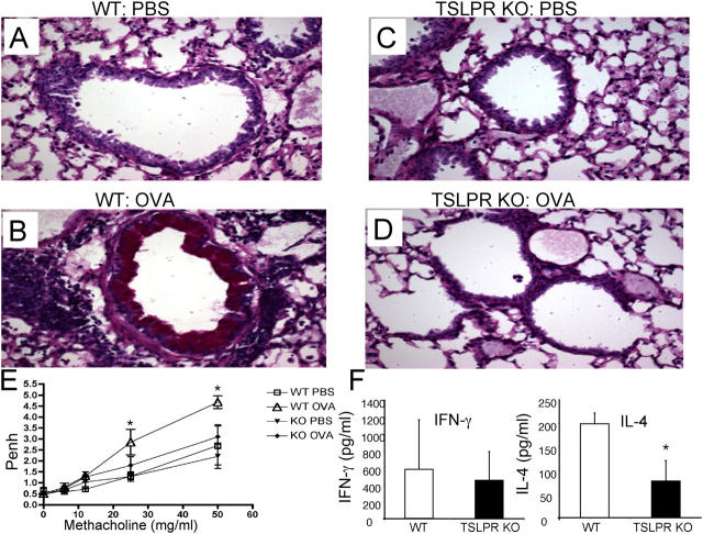
Figure 4.
TSLPR KO mice fail to mount an inflammatory response. (A–D) Periodic acid-Schiff–stained lung tissue sections of BALB/c WT and TSLPR KO mice that were sensitized (i.p.) and challenged (i.t. and i.n.) with OVA or PBS (i.p.). There were no obvious differences in the lung morphology between WT (A) and TSLPR KO (C) animals that were exposed to PBS. WT mice that received OVA displayed perivascular inflammation, peribronchiolar cuffing, and goblet cell hyperplasia (B), whereas TSLPR KO mice that were treated with OVA showed no obvious inflammation (D). (E) WT and TSLPR KO animals treated as shown above were tested for their airway hypersensitivity response using methacholine (0, 6, 12, 25, and 50 mg/ml). Unlike WT animals, TSLPR KO mice immunized and sensitized with OVA showed similar results to PBS control mice (six to seven animals per group, shown is one representative experiment out of three). *Statistical significance (P < 0.05) between the indicated dose and the 0 mg/ml. (F) Splenocytes from immunized WT and TSLPR KO mice were cultured in vitro with 200 μg/ml OVA for 3 d. Supernatants were examined for IFN-γ and IL-4 levels. The levels of IL-4 were significantly lower in the TSLPR KO animals versus the WT animals, whereas no statistical significance was observed for IFN-γ. The experiment was done four times with four mice in each experimental group in each experiment. *Statistical significance (P < 0.05).

