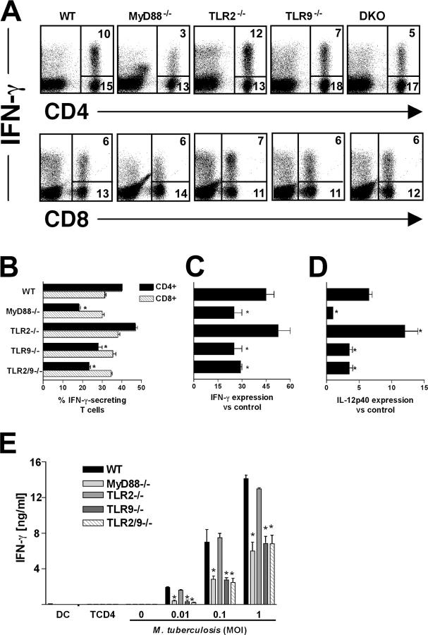
Figure 6.
Influence of TLR9 on the generation of IFN-γ–producing CD4+ T cells and Th1-associated cytokines in M. tuberculosis–infected mice. (A) Lung cells isolated from day 30 infected mice were stimulated with anti-CD3 mAb, and intracellular IFN-γ production was determined by flow cytometry after gating lymphocyte populations by forward and side scatter parameters. The FACS profiles of anti-CD4– and anti-CD8–stained lymphocytes in A are from pooled cells from two mice and are representative of results from four animals per group. The majority (85–95%) of the IFN-γ plus CD4− cells shown in the dot plots in the top panel were determined to be CD8+ T cells (unpublished data). Based on its nonspecific staining with multiple antibodies, the CD4 dim IFN-γ+ population in the lung preparations from infected MyD88 KO mice is likely to represent dead cells, consistent with their abundance in sections of the same tissue (Fig. 5, B and G). Percentage of CD4+ and CD8+ T cells that stain positively for IFN-γ calculated from the experiments shown in A. Relative expression of mRNAs for IFN-γ (C) and IL-12p40 (D) determined in lungs at 30 d after infection. Results are mean ± SE of measurements from three animals. (E) Purified splenic CD4+ T cells from the same mice described in A were cocultured with BMDCs infected with different MOIs for 72 h. IFN-γ was assayed by ELISA in culture supernatants. The means ± SE of measurements from triplicate wells are presented. The experiment shown was performed twice with similar results. *, significantly different values (P < 0.05) between WT and KO cells.

