
Figure 3.
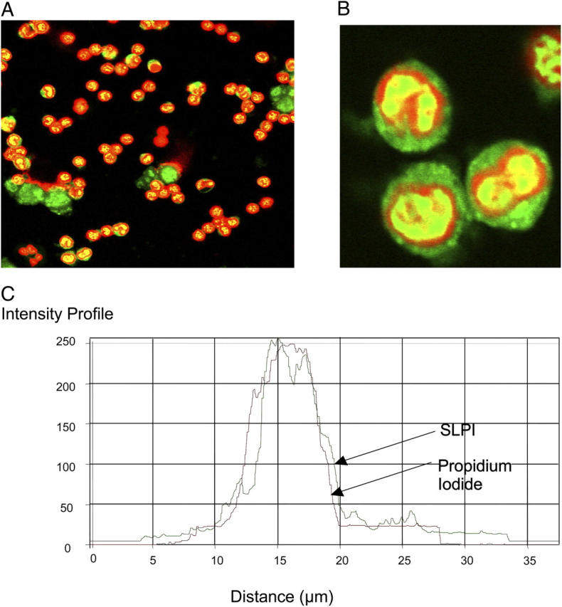
Nuclear import of SLPI-fluorescein in PBMs. 10 μg/ml SLPI-fluorescein (green) was incubated with monocytes for 1 h followed by counterstaining with propidium iodide. Red, nuclear stain. (A and B) Treated monocytes and (C) intensity profile. Red line, propidium iodide; green line, SLPI. (B) A magnification of the bottom left-hand quadrant of panel A.