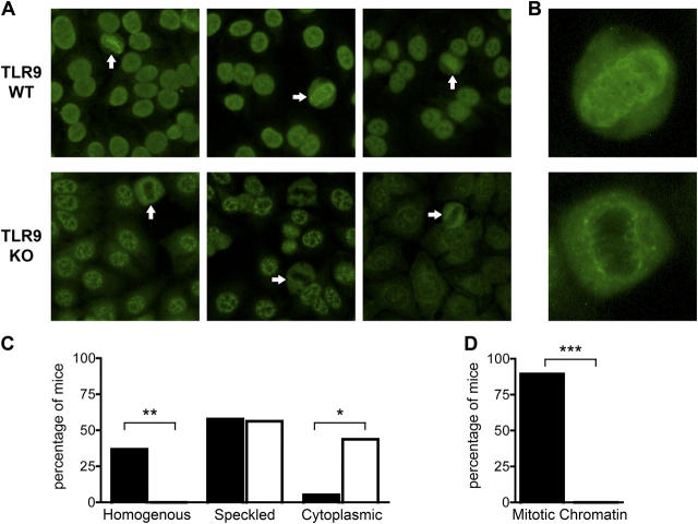
Figure 1.
TLR9-deficient sera lack anti-DNA and antichromatin staining patterns. (A) ANAs were determined in sera (1:200) from 20-wk-old F2 mice. TLR9+/+ sera are shown at the top (left, homogenous pattern; middle and right, speckled pattern); TLR9−/− sera are shown at the bottom (left and middle, speckled pattern; right, cytoplasmic pattern). Some TLR9+/+ sera had predominantly speckled patterns superimposed on faint homogenous staining (middle). White arrows indicate cells in metaphase that demonstrate positive (top, TLR9+/+) or negative (bottom, TLR9−/−) chromatin staining. Original magnification, 400. (B) Digitally enlarged images of metaphase cells with positive (top, TLR9+/+) or negative (bottom, TLR9−/−) chromatin staining. (C) Serum ANAs were classified as nuclear homogenous, nuclear speckled, or cytoplasmic staining patterns. Black bars indicate TLR9+/+ sera (n = 19), and white bars indicate TLR9−/− sera (n = 16). (D) As in C, but serum ANAs were classified as either positive or negative for mitotic chromatin staining. *, P < 0.02; **, P < 0.01; and ***, P < 0.0001 by Fisher's exact test.

