
Figure 3.
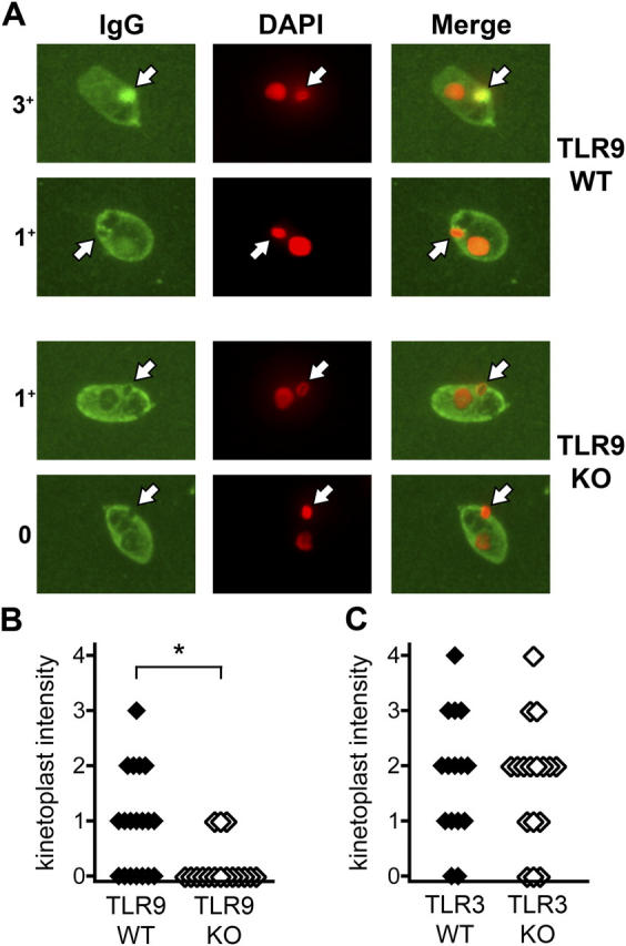
Reduced anti-dsDNA autoantibodies in TLR9−/− but not TLR3−/− mice. (A) Anti-dsDNA antibodies were detected by C. luciliae immunofluorescence. IgG antibodies to C. luciliae DNA are shown in green (left), and DAPI staining of DNA is shown in red (middle). White arrows indicate the kinetoplast. Specific anti-dsDNA antibodies are identified by colocalization of IgG and DAPI staining in the kinetoplast and appear in yellow (right). Representative TLR9+/+ sera are shown in the top two rows (intensity scores of 3+ and 1+), and TLR9−/− sera are shown in the bottom two rows (intensity scores of 1+ and 0). Original magnification, 1,000. (B and C) Specific anti-dsDNA staining of C. luciliae kinetoplasts was scored from 0 to 4 as in A for either TLR9+/+ (n = 19) and TLR9−/− (n = 16) sera (B), or TLR3+/+ (n = 15) and TLR3−/− (n = 17) sera (C). *, P < 0.02 by the Mann-Whitney U test.