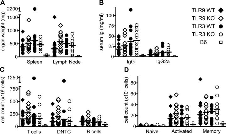
Figure 5.
Lymphadenopathy, hypergammaglobulinemia, and lymphocyte accumulation in TLR-deficient mice. TLR9+/+ (n = 19), TLR9−/− (n = 16), TLR3+/+ (n = 15), and TLR3−/− (n = 17) mice were killed at 20 wk of age and assessed for evidence of aberrant immune activation; nonautoimmune C57BL/6 control mice (n = 4) were killed at 26 wk of age. (A) Spleens and the two largest axillary lymph nodes were removed and weighed. (B) Total serum IgG and IgG2a were determined. (C) Splenocyte subsets were enumerated by FACS analysis for T cells (Thy1.2+), DNTC (CD4−/CD8− double-negative T cells), and B cells (CD22+). (D) Splenic CD4+ T cells were classified as either naive (CD44− CD62L+), activated (CD44+ CD62L+), or memory (CD44+ CD62L−) phenotype. The analysis in D was performed on 12 TLR3+/+ and 12 TLR3−/− mice. Horizontal lines represent mean values.

