
Figure 3.
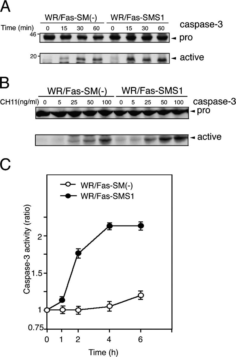
Fas-mediated caspase-3 activation in WR/Fas-SM(−) and WR/Fas-SMS1 cells. Time kinetics (A) and dose dependency (B) of caspase-3 activation by Western blot analysis. Cells were stimulated with 50 ng/ml CH11 for the indicated time (A), or stimulated with the indicated concentration of CH11 for 15 min (B). Total cell lysates were analyzed by Western blot using mouse mAb to caspase-3. The arrows indicate the bands corresponding to 32 kD for procaspase-3 (pro) and 17 kD for the active caspase (active). (C) Cells were stimulated with 50 ng/ml CH11 for the indicated time, and caspase-3 activities were measured in extracts of cell lysates using colorimetric assay kits. Each experiment was done in triplicate. Data are representative of five independent experiments. Error bars represent SEM.