
Figure 3.
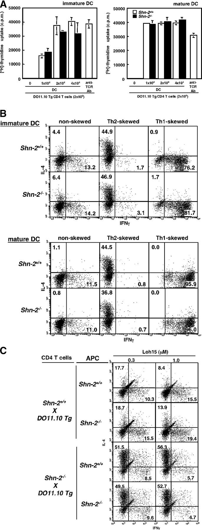
The function of Shn-2−/− APCs. (A) OVA-pulsed immature or mature BMDCs were cocultured with DO11.10 Tg CD4 T cells for 72 h. [3H]thymidine (37 kBq/well) was added to the stimulation culture for the last 16 h. The results (mean and standard deviation) of [3H]thymidine incorporation are shown. Three independent experiments were performed with similar results. (B) OVA-pulsed immature or mature BMDCs (2 × 10⁴ cells) were cocultured with DO11.10 Tg naive (CD44low) CD4 T cells (2 × 10⁵ cells) under nonskewed, Th2 cell–skewed, and Th1 cell–skewed conditions for 5 d. Intracellular staining profiles (IFN-γ/IL-4) of the cultured cells are shown with percentages of the cells in each quadrant. Three independent experiments were performed with similar results. (C) Naive (CD44low) CD4 T cells (1.5 × 10⁴ cells) were purified from Shn-2−/−× DO11.10 Tg mice by cell sorting and stimulated with indicated doses of specific OVA peptides (Loh15: 0.1 μM) and irradiated CD4− spleen cells (105 cells) from non-DO11.10 Tg Shn-2+/+or Shn-2−/−mice. Two independent experiments were performed with similar results.