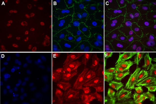
Figure 6.
Immunofluorescence localization of LSP1 in cultured human endothelial cells. HUVECs cultured on coverslips were double stained for LSP1 (red; A, C, E, and F) and VE-cadherin (green; B and C). (B–D) Cells were also counterstained for DAPI (blue). (C) The overlaid image of A and B. (D) Secondary Ab alone (Texas red) with DAPI staining. (E) The enhanced image (by increasing the contrast) of LSP1 staining pattern. (F) The enhanced image of LSP1 (from E) overlaid with phalloidin (green). Magnification, 400.

