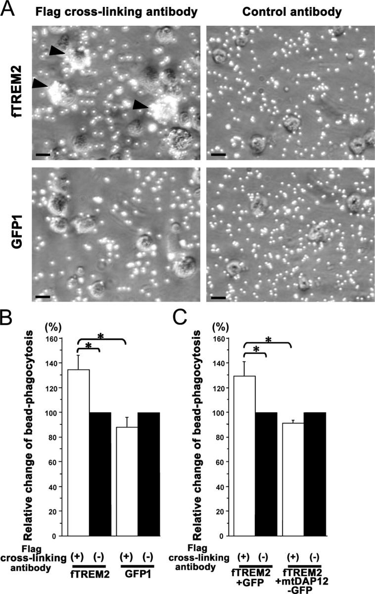
Figure 4.

Increased bead phagocytosis after stimulation of TREM2. (A) Primary murine microglia were transduced either with fTREM2 vector (fTREM2) or GFP1 control vector. Microglia were cultured on dishes coated with antibodies directed against the Flag epitope to cross-link the fTREM2 or control antibodies. Microglia were stimulated with antibodies for 24 h. Microsphere beads were then added for 1 h. Phase contrast images are shown, demonstrating visible phagocytosis of microshpere beads after stimulation of fTREM2-transduced microglia by cross-linking antibodies. Bars, 10 μm. (B) Phagocytosis was quantified by flow cytometry. The relative change in bead phagocytosis after fTREM2 stimulation was compared with nonstimulated bead phagocytosis. Microglia transduced with fTREM2 showed a significant increase in bead phagocytosis after cross-link stimulation, whereas no significant change in bead phagocytosis was observed in GFP1-transduced microglia after cross-link stimulation. Data are presented as mean ± SEM of at least three independent experiments. *, P < 0.05; Mann-Whitney U test. (C) The relative change in bead phagocytosis after fTREM2 stimulation compared with the nonstimulated situation by flow cytometry. Microglia was transduced with fTREM2 plus GIPγ or fTREM2 plus mutant DAP12 (mtDAP12-GFP). The mtDAP12 transduction prevented the increase in bead phagocytosis after fTREM2 stimulation. Data are presented as mean ± SEM of at least three independent experiments. *, P < 0.01; Mann-Whitney U test.
