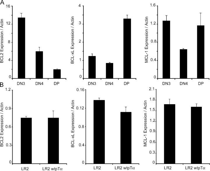
Figure 2.
BCL-2, BCL-xL, and MCL-1 are not induced by pre-TCR signaling. (A) Quantitative real-time RT-PCR analysis of BCL-2, MCL-1, and BCL-xL expression in the indicated thymocyte subsets. (B) Quantitative RT-PCR analysis of gene expression in the LR2 and LR2wtpTα cell lines. Actin expression was used to normalize gene expression.

