Figure 1.
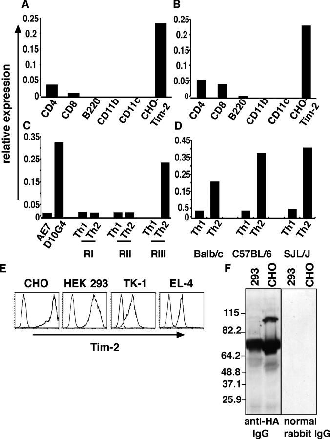
Tim-2 is up-regulated in Th2 cells. The expression pattern of Tim-2 was examined in various cell populations using real-time quantitative RT-PCR. Tim-2 expression was examined and expressed relative to GAPDH expression. (A) Ex vivo SJL CD4+, CD8+, B220+, CD11b+, and CD11c+ cells were purified from spleen cells. RNA was extracted and cDNA was generated for real-time quantitative PCR. (B) Ex vivo SJL CD4+, CD8+, B220+, CD11b+, and CD11c+ cells were purified from spleen cells. CD4+ and CD8+ cells were activated with plate-bound anti-CD3/anti-CD28; B220+, CD11b+, and CD11c+ were activated by LPS and IFN-γ. 48 h after activation, RNA was extracted and cDNA was generated for real-time quantitative PCR. (C) D011.10 CD4+ T cells were stimulated with OVA-peptide and APCs, in vitro under Th1 or Th2 polarizing conditions. At the end of each round of stimulation, RNA was extracted and cDNA was generated from Th1 and Th2 cells. (D) CD4+ T cells from SJL, C57BL/6, and BALB/c mice were stimulated with plate-bound anti-CD3/anti-CD28 in vitro under Th1 or Th2 polarizing conditions. At the end of the first round of stimulation, RNA was extracted and cDNA was generated from Th1 and Th2 cells. (E) Tim-2 is expressed as a cell surface protein. T cell (EL-4 and TK-1) and non–T cell (CHO and 293) transfectants were stained with biotinylated anti-HA antibody, and streptavidin-PE as a secondary detection reagent. Cells were analyzed by flow cytometry for surface expression of Tim-2. Solid line represents the staining of Tim-2 transfectants and the dotted line, empty vector. (F) Tim-2 expression by Western blot. Lysates from Tim-2–transfected CHO and HEK-293 cells were run on a 10% SDS-PAGE gel. Polyvinyldifluoride membranes were probed with anti-HA antibody or control normal rabbit IgG, followed by an HRP-conjugated anti–rabbit secondary antibody.

