
Figure 2.
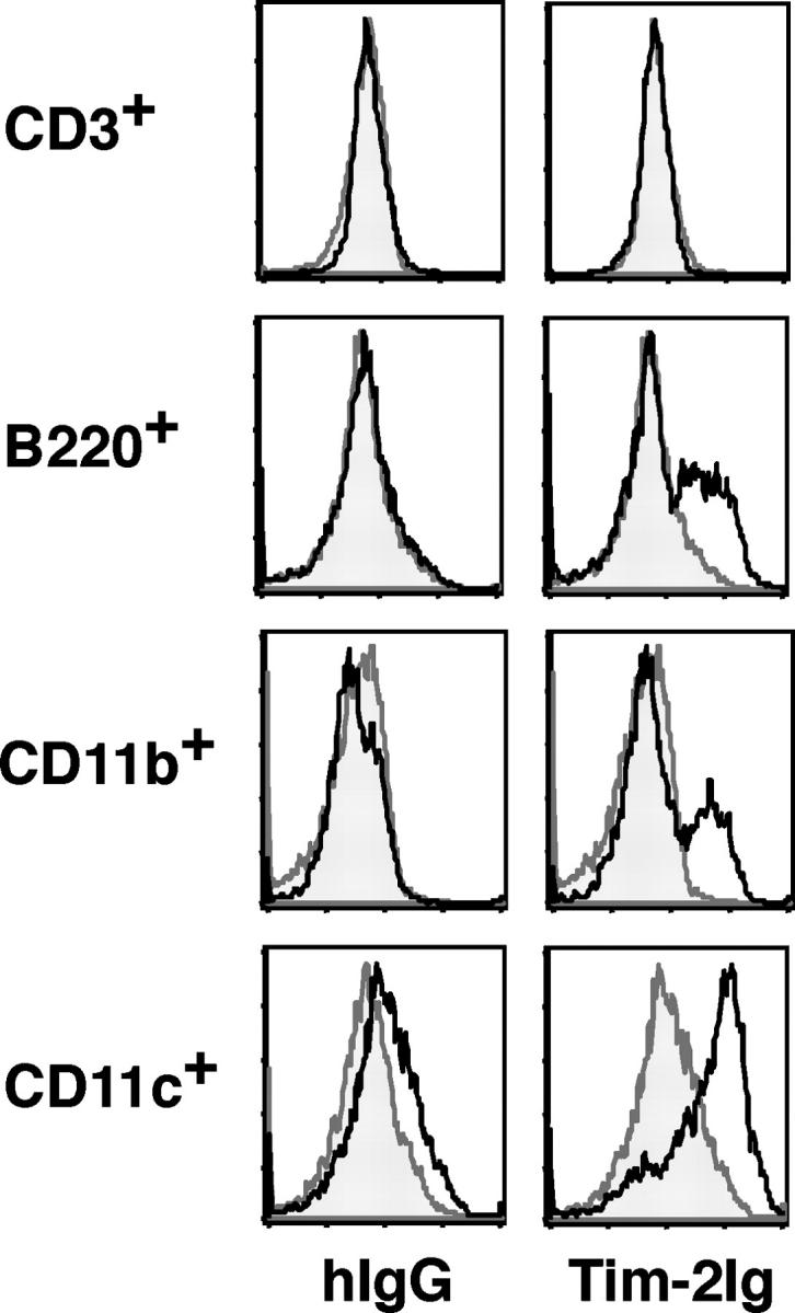
Tim-2 ligand is expressed on activated APCs. To determine the cell populations expressing Tim-2 ligands, we stained various cell populations with biotinylated Tim-2 Ig as follows: ex vivo BALB/c CD3+, B220+, CD11b+, or CD11c+ cells. Spleen cells from BALB/c mice were activated with conalbumin A (CD3+) or LPS and IFN-γ (B220+, CD11b+ and CD11c+), were column purified using negative selection (CD3+) or positive selection (B220+, CD11b+, and CD11c+) and stained for Tim-2 ligand expression with biotinylated Tim-2 Ig (solid black line), or biotinylated hIgG (shaded curve) as a control, followed by streptavidin-PE as a secondary reagent for detection.