Figure 2.
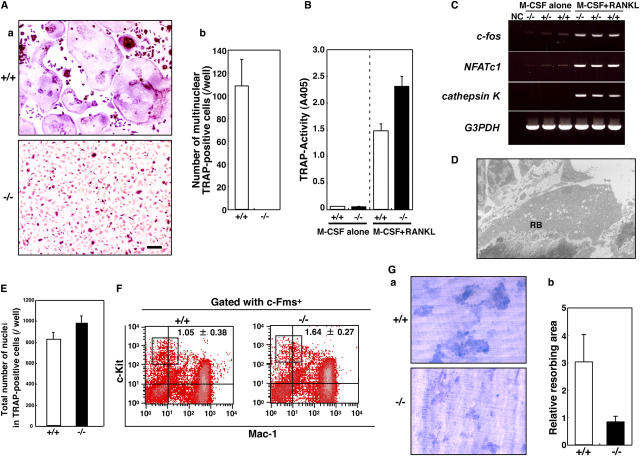
Lack of multinucleation in DC-STAMP−/−osteoclasts. (A) TRAP expression was induced, but multinucleation was completely abrogated in DC-STAMP−/− osteoclasts. TRAP staining (a) and the number of multinuclear TRAP-positive cells (b) are shown. Values represent SD. Bar, 50 μm. (B) TRAP solution assay of macrophages (M-CSF alone) and osteoclasts (M-CSF + RANKL). (C) Expression of c-fos, NFATc1, or cathepsin K in macrophages (M-CSF alone) or osteoclasts (M-CSF + RANKL) derived from DC-STAMP+/+, DC-STAMP+/−, or DC-STAMP−/− mice was analyzed by RT-PCR. NC, no template control. (D) Ruffled border formation was detected in osteoclasts of DC-STAMP−/− tibial sections under electron microscopy. RB, ruffled border. (E) Total number of nuclei in cultured osteoclasts. Values represent SD. (F) The percent frequency of a population of osteoclast precursor cells (boxes, c-Fms+c-Kit+Mac-1low) is shown as the mean ± SD. (G) Resorbing lacunae were visualized by toluidine blue O staining (a), and relative resorbing areas were scored (b). Values represent SD.

