Figure 1.
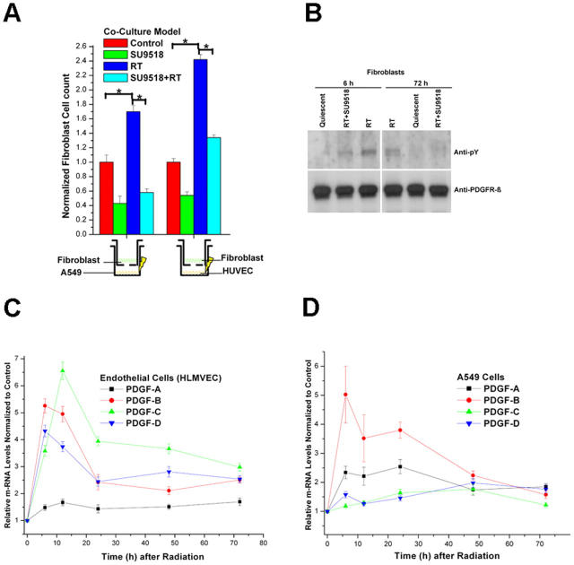
Radiation-induced paracrine activation of fibroblasts in a coculture system. (A) Fibroblast proliferation induced by exposure to coculture medium (Control) or by prior 10 Gy irradiation of HUVECs or A549 cells in the absence (RT) or presence of SU9518 (SU9518+RT) in the fibroblast medium. Mean ± SD. *, P < 0.05. (B) Phosphorylation status (anti-phosphorylated tyrosine antibody, anti-pY) of PDGFRβ in quiescent fibroblasts, fibroblasts exposed to medium from 10 Gy irradiated endothelial cells (6 and 72 h after radiation, RT), or with additional exposure to PDGF RTKI, SU9518 (RT+SU). Equal loading of lanes was demonstrated with anti-PDGFRβ. (C) Real-time quantitative RT-PCR of PDGF-A, PDGF-B, PDGF-C, and PDGF-D isoforms at 6, 12, 24, 48, and 72 h after 10 Gy irradiation of HLMVECs and A549 cells. Data are means ± SD from at least three independent measurements and show relative expression levels compared with the nonirradiated control cells at each time point.

