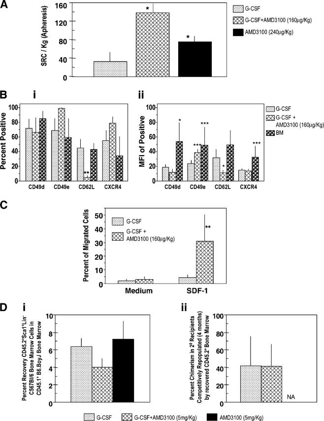
Figure 5.
Influence of AMD3100, G-CSF, and the combination of G-CSF plus AMD3100 on mobilization of NOD-SCID SRCs from normal human volunteers (apheresis samples), surface expression of adhesion molecules and chemotaxis of CD34+ cells, and homing of mobilized murine Sca1+Lin− cells. (A) SRCs per kg in apheresis samples from G-CSF– (n = 3), G-CSF + AMD3100- (160 μg/kg; n = 3), and AMD3100- (240 μg/kg; n = 4) mobilized circulating blood. Each set of test samples was assayed simultaneously in limiting dilutions in conditioned NOD-SCID mice. For every sample, four different cell concentrations were used and four mice were transplanted with each cell concentration. Mice were assayed for chimerism 8 wk later; those that demonstrated >0.2% chimerism (total CD45+ cells in BM) were considered to be positive. Percentage of negative mice were used to calculate SRC frequencies. (B, i) Expression of CD49d (VLA-4), CD49e (VLA-5), CD26L (L-selectin), and CXCR4 on mobilized CD34+ cells (mean ± 1SEM of percent positive cells of five to seven different G-CSF samples, three AMD3100 plus G-CSF samples, and four BM samples). (B, ii) Mean fluorescent intensity (MFI) of positive samples. Same number of samples evaluated as in B, i except G-CSF group has a different number (n =7). (C) CD34+ cells isolated from G-CSF (n = 6) and G-CSF plus AMD3100 (n = 3) mobilized peripheral blood were assessed for chemotaxis to SDF-1/CXCL12 (100 ng/ml) and results expressed as percentage of migrated cells. Data for each sample were collected in duplicate. (D, i) Homing of CD45.2+ Sca1+Lin− BM cells from C57Bl/6 mice into lethally irradiated CD45.1+ B6.BoyJ BM from samples mobilized with G-CSF (2 times/d for 4 d as in Fig. 2B; n = 3), G-CSF + AMD3100 (5 mg/kg; n = 4), and AMD3100 (5 mg/kg; n = 3). Homing was assessed as in Materials and methods. (D, ii) Competitive repopulation of CD45.2+ BM cells recovered after homing shown in D, i. Results of chimerism are after 4 mo in 2° irradiated B6.BoyJ recipients. For A–D, significant differences compared with G-CSF: *P < 0.001; **P < 0.01; ***P < 0.05; all other values are P > 0.05. NA, data not depicted.

