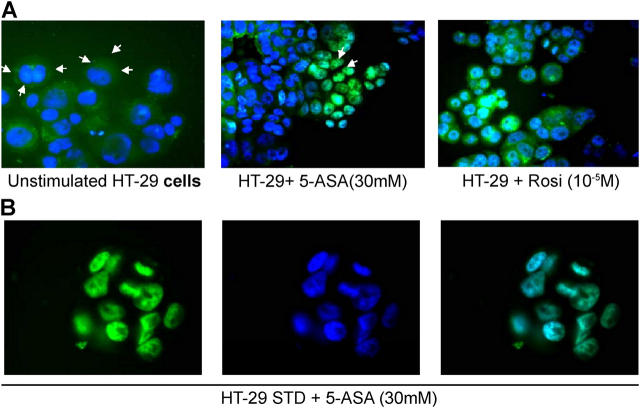
Figure 4.
Intracellular localization of PPAR-γ in epithelial cells. (A) HT-29 STD cells transfected with GFP-tagged PPAR-γ were incubated in the presence of medium alone (unstimulated cells), 5-ASA (30 mM), or rosiglitazone (rosi, 10−5 M) for 24 h. Nuclear staining in blue was performed with Hoescht 33342 solution. The intracellular distribution of the fluorescent tags was examined under a fluorescence microscope. (B) HT-29 STD cells transfected with GFP-tagged PPAR-γ incubated in the presence of 5-ASA (30 mM) for 24 h. Green color represents PPAR-γ (left); blue color represents the nucleus (middle); merged green and blue pictures illustrate the translocation of PPAR-γ into the cell nucleus (right).

