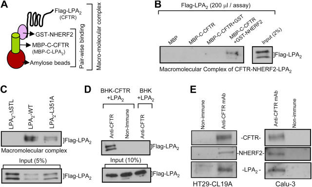
Figure 6.
LPA2 forms a macromolecular complex with CFTR mediated through NHERF2. (A) Pictorial representation of the macromolecular complex assay (see Materials and methods for details). (B) Macromolecular complex of MBP-C–CFTR, GST–NHERF2, and Flag-tagged LPA2. (C) Macromolecular complex of MBP-C–CFTR, GST–NHERF2, and Flag-tagged LPA2, LPA2–ΔSTL, or LPA2–L351A. (D) BHK or BHK–CFTR cells transiently transfected with Flag-LPA2 were cross-linked using 1 mM DSP and coimmunoprecipitated using anti-CFTR antibody (NBD-R) and probed for LPA2. (E) Cell lysates from HT29-CL19A (left) and Calu-3 (right) were coimmunoprecipitated using anti-CFTR antibody (R1104) and probed for NHERF2 and LPA2. LPA2 blot was visualized using SuperSignal West Femto Maximum Sensitivity Substrate because of the weak signal (Pierce Chemical Co.).

