
Figure 6.
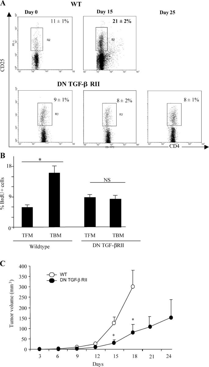
TGF-β signaling is critical for T reg cell proliferation and tumor escape. (A) Flow cytometry analyses of CD3+CD4+CD25+ cells isolated from the inguinal LNs of WT and DN TGF-βRII mice. The animals were either tumor free (0), or bore 15- or 25-d-old tumors. Data from one representative animal in each group is shown. Percentages of CD25 expression among CD3+CD4+ cells are shown. Values represent the mean ± SEM (n = 6). (B) BrdU incorporation into CD3+CD4+CD25+-gated T cells isolated from the inguinal LNs of WT and DN TGF-βRII mice. Animals were either tumor free or bore 15-d-old tumors, as indicated by the symbols, and the frequency of BrdU+ cells was determined. Values represent the mean ± SEM (n = 6). One representative experiment out of two is shown. The asterisk indicates statistical significance (P < 0.05) using the Student's t test. (C) Influence of the DN TGF-βRII transgene on PROb tumor growth. WT (open circles) and transgenic DN TGF-βRII mice (closed circles) were challenged s.c. with B16F10 cells, and tumor growth was monitored biweekly. One representative out of two experiments including six mice per group following the mean tumor volume ± SEM is depicted. The asterisk indicates statistical significance (P = 0.001) at 95% confidence using the Student's t test.