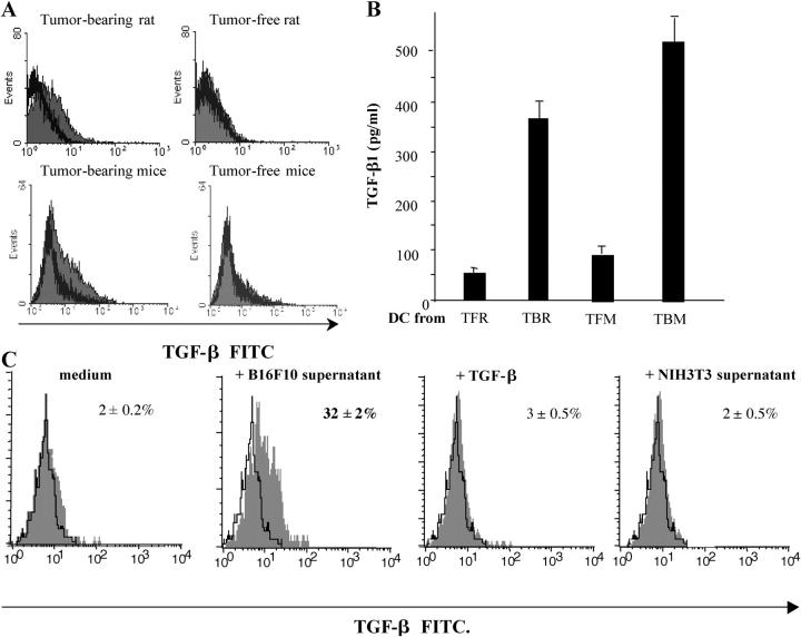
Figure 7.
Tumor cells license IMDCs to produce TGF-β. (A) Intracellular staining of IMDCs from tumor-free or tumor-bearing rats and mice. CD11b+ DCs isolated from the spleen of TBR and TFR or CD11c+ IMDCs from DLNs of TFM or TBM were permeabilized and stained with a polyclonal anti–TGF-β antibody. Shaded histogram, anti–TGF-β; bold line histogram, isotype control. Note that no positive staining was obtained with nonpermeabilized cells (not depicted). (B) TGF-β secretion by IMDCs. IMDCs purified as in A were cultured for 48 h in serum-free medium and TGF-β1 levels in the supernatants were assessed by ELISA. Values represent the mean ± SEM (n = 3). (C) Induction of TGF-β production in mouse splenic IMDCs by tumor supernatants. CD11c+ DCs from inguinal LNs of TFM were cultured for 24 h in the absence or presence of B16F10 or NIH 3T3 culture supernatants or 10 ng/ml TGF-β1. Shaded histogram, test tube; open histogram, isotype control. At 24 h, TGF-β–expressing cells were identified by FACS analysis after permeabilization. The percentages of TGF-β expression cells are given. Data representative of two independent experiments are shown.

