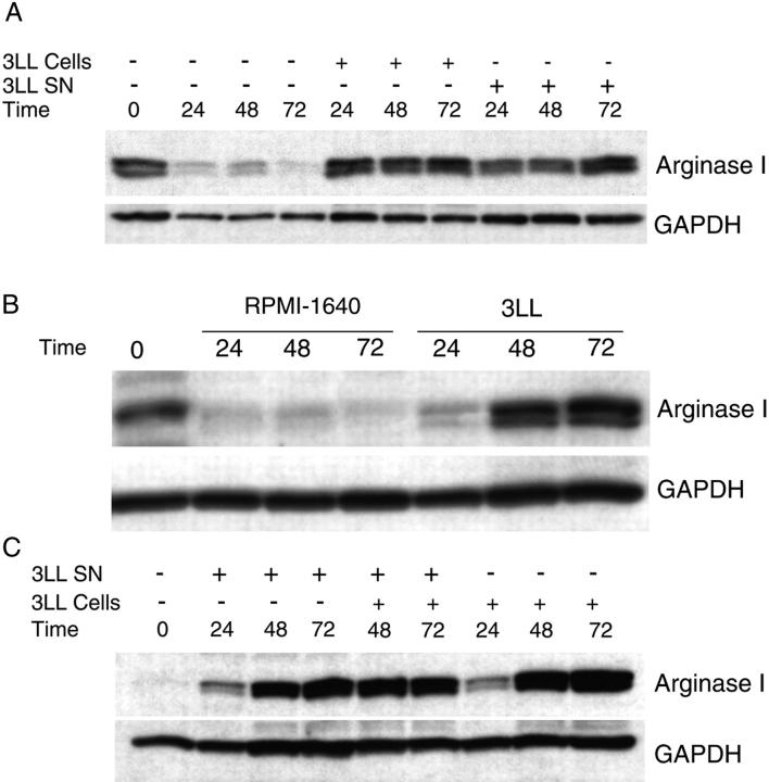
Figure 2.
Arginase I expression in MSCs is induced by tumor-derived soluble factors. (A) MSCs (2 × 10⁶) isolated from 3LL tumors were cultured alone or cocultured in transwells with 2 × 10⁶ 3LL cells or 3LL supernatants. Arginase I expression was tested via Western blot analysis at 24, 48, and 72 h. (B) MSCs (2 × 10⁶) were cultured for 24 h in regular RPMI-1640, which results in the loss of arginase 1 expression. They were then cultured in RPMI-1640 alone or cocultured in transwells with 3LL cells (2 × 10⁶). Arginase I expression was tested at 24, 48, or 72 h afterward via Western blot analysis. (C) Peritoneal macrophages (2 × 10⁶) from normal mice were cocultured in transwells with 3LL cells (2 × 10⁶) or 3LL supernatants. Arginase I expression was tested via Western blot analysis. Results shown are representative of 3 experiments.

