
Figure 1.
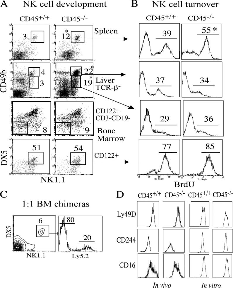
The absence of CD45 alters NK cell development. (A) Spleen and liver from CD45−/− mice were analyzed for NK1.1+CD49b+ NK cells by flow cytometry. NK cell precursors from bone marrow were identified as CD122+NK1.1−DX5−CD3−CD19−. Data are representative of 12 mice or 6 mice for precursor analysis with percentages displayed next to each population. (B) Mice were given drinking water containing BrdU for 2 wk. Cells were stained with the same antibodies as in A with the addition anti-BrdU and analyzed by flow cytometry. Percentages of divided cells (BrdU+) are shown above the each population, with CD45−/− spleen-derived NK cells having a greater percentage of dividing cells (*, P = 0.008). Data are representative of three independent experiments. (C) Hematopoietic chimeras were generated by injecting irradiated Ly5.1 mice with equal amounts of control (Ly5.2) and CD45−/− bone marrow. The resulting NK1.1+DX5+ population was analyzed for expression of Ly5.2 by flow cytometry to deduce donor origin. (D) The level of Ly49D, CD244, and CD16 protein expression on the surface of freshly isolated and IL-15–expanded NK1.1+CD49b+ cells was analyzed by flow cytometry. Data shown for C and D are representative of six independent experiments.