
Figure 2.
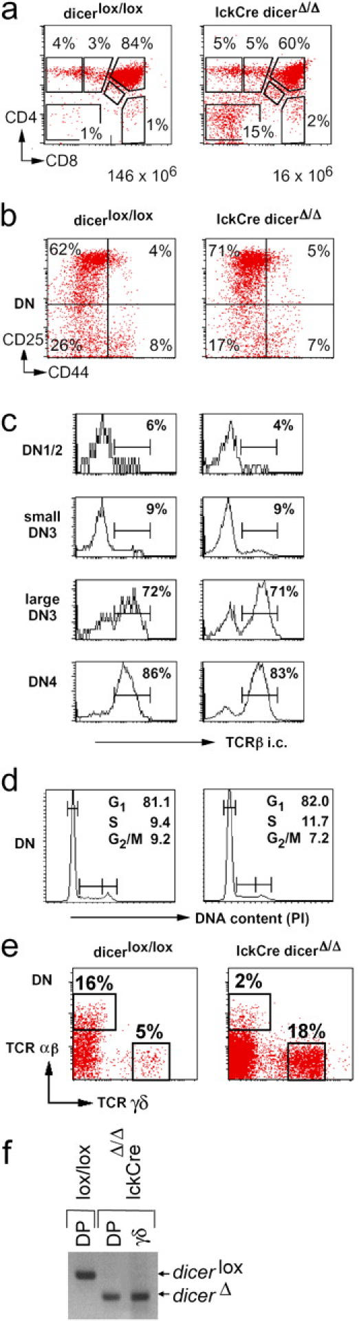
Reduced cellularity of lckCre Dicer Δ/Δ thymi, but no developmental block at the DN stage. (a) Thymocyte numbers and subset distribution defined by CD4 and CD8 expression in Dicer lox/lox and lckCre Dicer Δ/Δ littermates. The representation of thymocyte subsets and the total number of thymocytes are indicated. Note reduced cellularity in lckCre Dicer Δ/Δ thymi. (b) Expression of CD44 and CD25 on DN cells indicates normal DN subset distribution in lckCre Dicer Δ/Δ thymocytes. (c) Intracellular staining of DN thymocyte subsets indicates normal TCR-β expression in lckCre Dicer Δ/Δ DN thymocytes. (d) DNA content as assessed by propidium iodide (PI) staining indicates that lckCre Dicer Δ/Δ DN thymocytes proliferate normally. (e) TCR γδ-expressing cells are overrepresented in the absence of Dicer. (f) Genomic PCR shows that Dicer deletion is comparable, and virtually complete, in lckCre Dicer Δ/Δ γδ-expressing thymocytes.