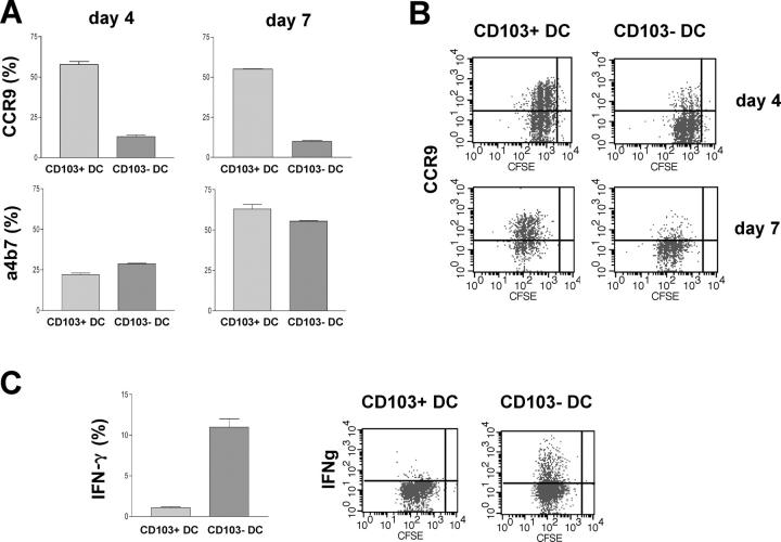
Figure 6.
Functional characteristics of CD11chigh CD103+ cells. MACS-sorted, CFSE-labeled CD4+ cells from DO11.10 SCID donors were incubated with FACS-sorted CD103+CD11chigh or CD103−CD11chigh DCs from MLN of Balb/c donors and OVA peptide. T cells were analyzed by FACS for expression of CCR9 and α4β7 at day 4 of culture and, after a further 3 d of expansion in IL-2. At day 7 of culture, T cells were restimulated overnight with platebound anti-CD3 mAb before intracellular cytokines were assessed. (A) Mean percentage ± SD of T cells expressing CCR9 and α4β7. Data from one of two independent experiments. (B) Representative staining of CCR9 on T cells. (C) Mean percentage ± SD of T cells producing IFN-γ and representative stainings. Data from one of two independent experiments.

