
Figure 2.
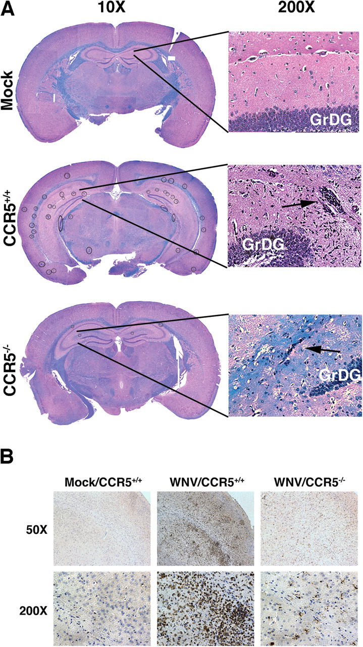
West Nile virus induces multifocal encephalitis in C57BL/6 mice. (A) Histologic analysis. Brains of mock- and WNV-infected CCR5+/+ and CCR5−/− mice harvested at day 12 after infection were fixed in formalin and embedded in paraffin. Coronal sections 6-μm thick were stained with hematoxylin and eosin to visualize cells and luxol fast blue to stain myelin tracts blue. Multiple cellular infiltrates were observed in the infected mice (circled in the 10× image). Typical infiltrates are shown at higher magnification in the hippocampus (arrows). GrDG, granule cell layer of the dentate gyrus. (B) Immunohistochemical analysis. Brain sections, prepared as in A from mock- and WNV-infected mice with the indicated CCR5 genotype, were stained with methyl green and anti–human CD3 mAb. Representative sections of the cortex are shown. CD3+ cells stain brown. Images in A and B are representative of five mock-infected brains, nine WNV-infected CCR5+/+ brains, and six WNV-infected CCR5−/− brains.